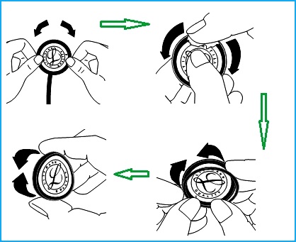
How to remove and
assemble the Diagram _

The stethoscope is most often used to listen to heart sounds and
breathing. There are two basic types of stethoscopes for respiration system
diagnostics of the human body.
Acoustic
Stethoscope
Acoustic stethoscopes are familiar to most people. Acoustic stethoscopes maintain their popularity and trust among doctors due to their longstanding use in the medical profession. Although it is taking digital stethoscopes time to catch up, they are rapidly gaining in popularity. However, acoustic stethoscopes are the most commonly used.
Acoustic stethoscopes are familiar to most people. Acoustic stethoscopes maintain their popularity and trust among doctors due to their longstanding use in the medical profession. Although it is taking digital stethoscopes time to catch up, they are rapidly gaining in popularity. However, acoustic stethoscopes are the most commonly used.
Electronic
Stethoscope
Electronic stethoscopes function in a similar way as acoustic stethoscopes, but the sound is converted to electrical signals which can then be amplified and processed for optimal listening. Because the sounds are transmitted electronically, an electronic stethoscope can be a wireless device, can be a recording device, and can provide noise reduction, signal enhancement, and both visual and audio output.
Electronic stethoscopes function in a similar way as acoustic stethoscopes, but the sound is converted to electrical signals which can then be amplified and processed for optimal listening. Because the sounds are transmitted electronically, an electronic stethoscope can be a wireless device, can be a recording device, and can provide noise reduction, signal enhancement, and both visual and audio output.
ADVANTAGES & DISADVANTAGES
> In Digital stethoscope Amplifiers are used which amplifies the low level signal.
> Data can be stored for further analysis and consultation.
> Heart signal is displayed on the LCD screen.
> In digital stethoscope battery replacement can be tedious.
> They are expensive.
> They suffer from interference when other electronic instruments like cell phones are around.
PRINCIPLE OF OPERATION
The stethoscope is composed of three major parts. The first part is the chest piece, tubes and headset. It consists of a shallow, bell-shaped piece and a clear, stiff diaphragm, which is connected to the metal earpieces by a flexible tube. The bell is used to pick up lower frequency sounds, and the diaphragm is used for higher frequency sounds. When the chest piece is placed on the skin, vibrations within in the body are amplified by either the bell or diaphragm. These acoustic pressure waves then travel up through the tubing to the earpieces and into the listener’s ears. Digital stethoscopes offer new opportunities for computerized analysis of heart sounds. In cardiac auscultation, an examiner uses a stethoscope to listen to these sounds, which provide important information about the body condition. When the heart sounds are displayed graphically, the methodology is known as phonocardiography. As heart sounds are non-stationary signals, it is important to study both their temporal and frequency contents. Recently, with the rapid development of computer hardware and digital signal processing, heart sounds could be recorded and analyzed easily. The majority of these researchers mainly emphasize on the characteristic extraction by frequency-analysis method. Others concern on how to extract the heart rate from a weeping infant through the heart sound signals, and improve signal-to-noise ratio by adaptive filtering way.
Removing the Diaphragm and Cleaning the
Chest-piece.The stethoscope is composed of three major parts. The first part is the chest piece, tubes and headset. It consists of a shallow, bell-shaped piece and a clear, stiff diaphragm, which is connected to the metal earpieces by a flexible tube. The bell is used to pick up lower frequency sounds, and the diaphragm is used for higher frequency sounds. When the chest piece is placed on the skin, vibrations within in the body are amplified by either the bell or diaphragm. These acoustic pressure waves then travel up through the tubing to the earpieces and into the listener’s ears. Digital stethoscopes offer new opportunities for computerized analysis of heart sounds. In cardiac auscultation, an examiner uses a stethoscope to listen to these sounds, which provide important information about the body condition. When the heart sounds are displayed graphically, the methodology is known as phonocardiography. As heart sounds are non-stationary signals, it is important to study both their temporal and frequency contents. Recently, with the rapid development of computer hardware and digital signal processing, heart sounds could be recorded and analyzed easily. The majority of these researchers mainly emphasize on the characteristic extraction by frequency-analysis method. Others concern on how to extract the heart rate from a weeping infant through the heart sound signals, and improve signal-to-noise ratio by adaptive filtering way.
With the diaphragm side up, grasp the rim with the thumbs and
index fingers of both hands and roll the rim off the edge of the chest-piece.
Remove the diaphragm from the rim and clean the parts in soapy water or wipe with alcohol. Chest-piece surfaces can be wiped with alcohol or soapy water.
Remove the diaphragm from the rim and clean the parts in soapy water or wipe with alcohol. Chest-piece surfaces can be wiped with alcohol or soapy water.
Dry all parts and surfaces thoroughly before reassembly.
Assembly and Replacement of the Tunable
Diaphragm
Using your fingertip, apply a small amount of talcum powder to the
inside surface of the flexible edge of the diaphragm.
This will facilitate assembly and maintain smooth and quiet low/high frequency alternation.
This will facilitate assembly and maintain smooth and quiet low/high frequency alternation.
Snap the diaphragm with your fingers to remove excess talc.
Insert the flexible edge of the diaphragm into the groove of the
rim. This is best accomplished by starting with the rim positioned above the
legible side of the diaphragm.
Visually examine the ring to ensure that the flexible edge is smoothly engaged inside the rim. If necessary, flex the assembly by pinching the outer rim edge. between the thumb and fingers.
Repeat this flexing procedure after rotating the assembly one quarter.
Visually examine the ring to ensure that the flexible edge is smoothly engaged inside the rim. If necessary, flex the assembly by pinching the outer rim edge. between the thumb and fingers.
Repeat this flexing procedure after rotating the assembly one quarter.

To attach the rim/diaphragm assembly to the chest-piece, engage
the groove of the rim (with the diaphragm attached) around the chest-piece at
one point and hold it in place with your thumbs.
Slowly roll the rim around and over the chest-piece edge using
both thumbs, moving in opposite directions around the chest-piece.
Visually inspect the edge where the diaphragm engages the rim for
uniform containment.
Minor adjustments may be made by slightly pulling and rolling the rim away from the diaphragm, allowing the diaphragm to slip into position.
Minor adjustments may be made by slightly pulling and rolling the rim away from the diaphragm, allowing the diaphragm to slip into position.
Avoid extreme heat, cold, solvents, and oils.
Do not immerse your stethoscope in any liquid or subject it to
steam sterilization. If disinfection is required, the stethoscope may be wiped
with a 70% isopropyl alcohol solution. Wipe and clean with alcohol or soap
water.